Under the Microscope
Explore seven micrographs captured by members of the lab. Select an image to see it in full.
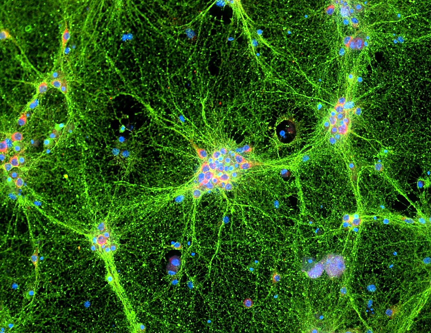
Neurons exposed to extracellular vesicles
Neurons exposed to a therapeutic extracellular vesicle (EV) treatment. Fluorescence labels show neurons (green) and EVs (red).
Microscopy by Dr. Lawrence
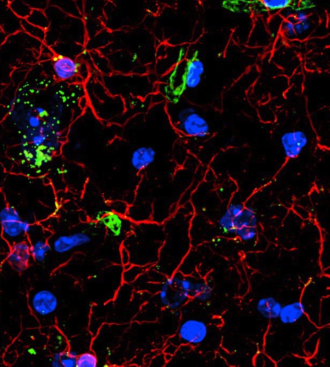
Somatosensory cortex after EV therapy
Somatosensory cortex from a mouse model of Alzheimer’s disease treated with therapeutic engineered extracellular vesicles (EVs). Fluorescence labels show microglia/macrophages (Iba1, red), lysosomes (CD68, green), and cell nuclei (DAPI, blue).
Microscopy by Dr. Patel
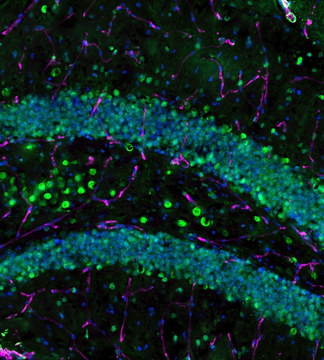
Dentate gyrus after cell therapy
Dentate gyrus in the hippocampus of a mouse model of Alzheimer’s disease treated with cell therapy. Fluorescence labels show smooth muscle actin (SMA, green), vascular endothelial cells (CD31, magenta), and cell nuclei (DAPI, blue).
Microscopy by Dr. Areiza-Mazo
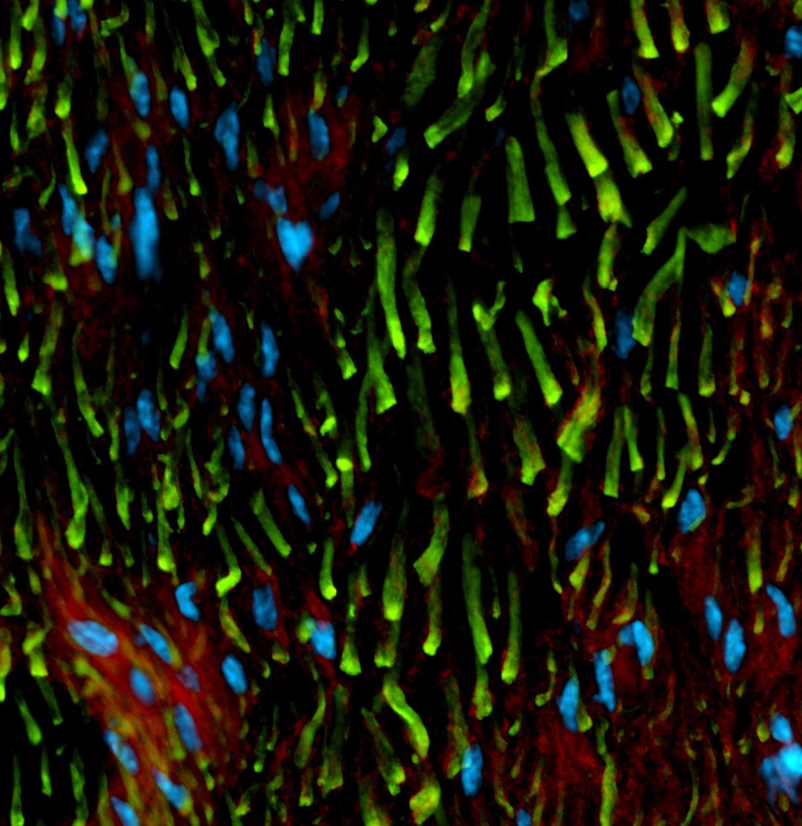
Gene delivery after sciatic nerve crush
Sciatic nerve from a mouse treated after a crush injury with vasculogenic non-viral gene therapy delivered by Tissue Nano-Transfection (TNT). Fluorescence labels show nerve fibers (neurofilament, green), blood vessels (red), and cell nuclei (DAPI, blue).
Microscopy by Dr. Moore
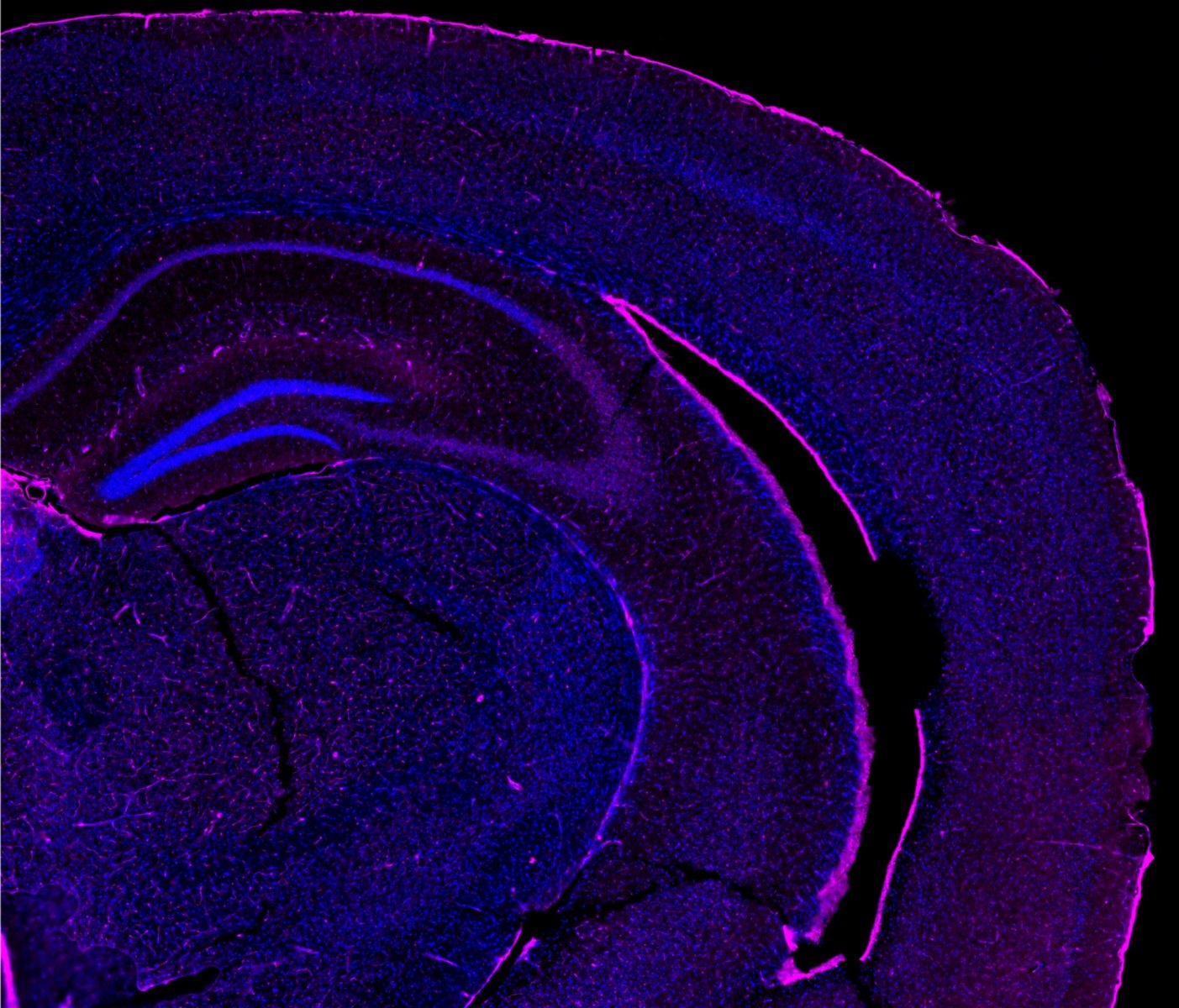
Brain section from an Alzheimer’s mouse model
Brain section from a mouse model of Alzheimer’s disease treated with a non-viral cell therapy aimed at repairing damaged or diseased brain tissue. Fluorescence labels show neurons and their processes (Tuj1, red) and cell nuclei (DAPI, blue).
Microscopy by Dr. Panić
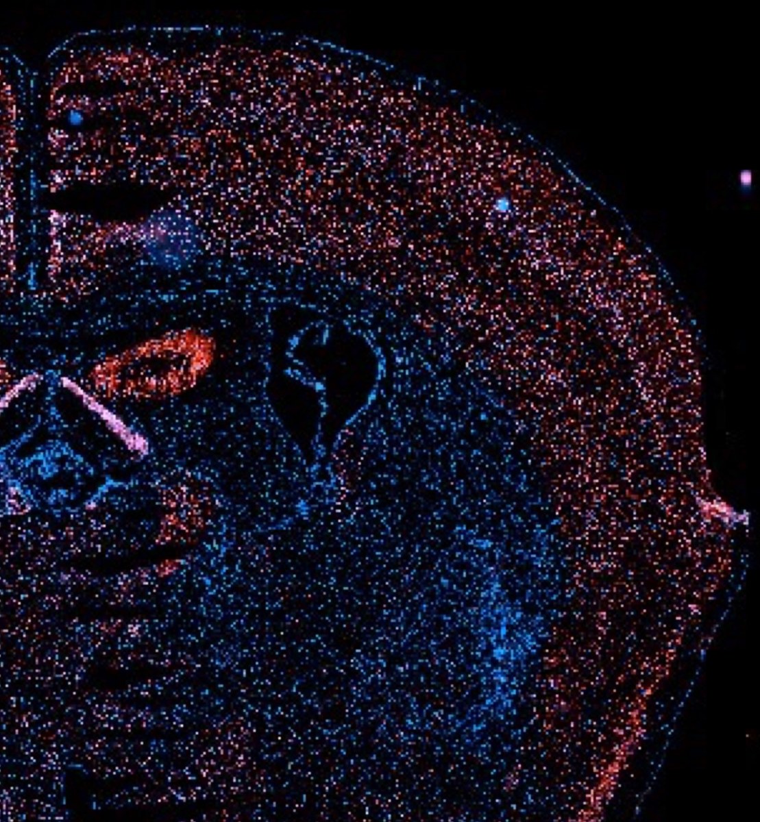
Cell therapy after stroke
Brain section from a mouse treated with vasculogenic cell therapy after stroke. Fluorescence labels show neurons (NeuN, red) and cell nuclei (DAPI, blue).
Microscopy by Dr. Lemmerman
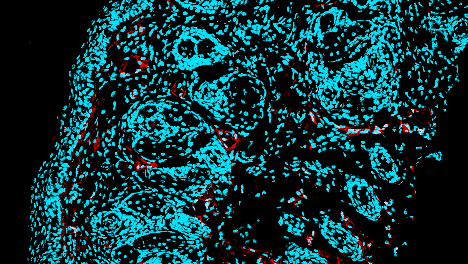
Blood vessel growth in human skin
Human skin showing blood vessel growth after vasculogenic Tissue Nano-Transfection (TNT). Fluorescence labels show vascular endothelial cells (CD31, red) and cell nuclei (DAPI, cyan).
Microscopy by Ms. Chaharsoughiamin